|
Splenomegaly in
Patients with Sideropenic Anemias: Clinical and
Hematologic Significance
......................................................................................................................................................................
Safaa A. A. Khaled (1)
Gehan S. Seifeldein (2)
(1) MD, Department of Internal Medicine, Hematology
& BMT unit, Assiut University Hospital, Faculty
of Medicine, Assiut University, Assiut, Egypt.
(2) MD, Department of Diagnostic Radiology, Assiut
University Hospital, Faculty of Medicine, Assiut
University, Assiut, Egypt.
Correspondence:
Dr. Safaa A.A. Khaled
Department of Internal Medicine, Hematology &
BMT unit.
Assiut University Hospital, Assiut, Egypt.
Tel: 002 0882 413940.
Email: sa_ah_mh@yahoo.com;
safaakhaled2003@gmail.com
|
ABSTRACT
Background, Objectives:
Sideropenic
anemias (SAs) are a group of hypoproliferative
anemias characterized by hyposideremia.
Although they run an insidiously started
slowly progressive course, they are a pointer
for an underlying serious disease. Fortunately,
in most cases, management of SAs is available,
effective and relatively inexpensive. Splenomegaly
was reported in patients with SAs with variation
in Hackett's grading and hematological profile.
Etiopathogenesis of splenomegaly in SAs
was mainly explained as related to the underlying
pathologic process of anemia or as a component
of the rarely occurring Paterson-Kelly syndrome.
Apart from the etiopathogenesis of splenomegaly
of SAs it is still a fruitful point for
current research. The aim of the present
study was to assess splenomegaly in patients
with SAs in terms of frequency, clinical
and hematological profile of splenomegaly
in SAs. Another aim was to assess prognostic
significance and to assume etiopathogenesis
of splenomegaly in SAs.
Methods: A prospective study was
conducted on 83 patients with SAs and 25
normal sex and age matched healthy controls.
Patients' demographics, clinical and hematologic
data were collected through thorough history
and clinical examination. Splenomegaly was
assessed with clinical examination of the
study subjects and was graded with Hackett's
clinical grading, then confirmed with ultrasonographic
examination. Patients were treated as per
the published guidelines for treatment of
SAs. Those with splenomegaly were subjected
to a strict follow up plan.
Results and Conclusion: Analysis
of the collected data showed that splenomegaly
is of robust clinical and hematologic significance
in patients with SAs.
Key words: Sideropenic anemias, splenomegaly,
Clinical significance.
|
Sideropenic anemia is a hematologic
term referred to anemias with reduced serum iron
levels; the term includes iron deficiency anemia
(IDA), and anemia of chronic disease (ACD), or
a dimorphic anemia of IDA and ACD. SAs are the
most prevalent types of anemias worldwide, firstly
IDA and secondly ACD. (1-3)
In vitro and in vivo studies demonstrated reduced
serum iron in patients with chronic inflammatory
conditions, infections and malignancies. Inflammatory
cytokines such as interleukin-1, interleukin-6,
and tumor necrosis factor-alpha are the main trigger
for hypoferemia; other bone morphogenetic proteins
2, 4, 6, & 9 produce the same effect in patients
with malignancy. These effects were mediated through
hepcidin. (4-6)
The differentiation between IDA and ACD is quiet
difficult, nevertheless in ACD there is confounding
evidence of chronic infectious, inflammatory,
or malignant disease causing the anemia. Furthermore
in ACD the RBCs indices are usually normal (MCV
from 80- 100fl, MCHC from 32-36 gm/dl and RDW
12.0-14.6%), while in IDA all RBCs indices are
below normal except the RDW which is commonly
raised. Total iron binding capacity (TIBC) was
found raised in IDA and reduced in ACD, however
serum hepcidin levels were considered the most
important difference between IDA and ACD. Unfortunately,
laboratory assay of serum hepcidin is difficult,
expensive and not widely available. Soluble transferrin
receptor (sTfR) was found to be a good differential
test between IDA and ACD; it was found raised
in patients with IDA however standardization of
the test was difficult. (7-9)
Hepcidin is a hepatic protein that is found to
be raised in patients with ACD and reduced in
IDA. Inflammatory cytokines are the most important
triggers for hepcidin production. Hepcidin affects
iron homeostasis by inhibition of a divalent iron
transporter protein-1, that in turn hinders enteral
iron absorption; and blocking a ferroprotin that
inhibits release of iron from iron stores. Both
cause sideropenia and raised iron levels in the
reticulo-endothelial tissues. (9,10)
Splenomegaly was reported in patients with SAs;
in IDA splenomegaly was described with Paterson-Kelly
syndrome, and hypopituitarism whereas in ACD it
is a diagnostic feature of the underlying disease.
The classic triad of Paterson-Kelly syndrome is
retropharyngeal dysphagia, eosophageal web, and
iron deficiency anemia. (11-14)
This study was conducted to evaluate the frequency,
and clinical significance (diagnostic/prognostic)
of splenomegaly in patients with SAs, also to
assess the association between different grades
of splenomegaly and both clinical and hematological
profiles of patients.
2.1. Study design and subjects
A prospective longitudinal study was conducted
at the Department of Internal Medicine, Assiut
University Hospital over a period of 6 months.
Three groups of patients were enrolled in the
study, patients with IDA patients with ACD, and
another group of gender and age matched healthy
volunteers was included as controls. Patients
were recruited among those who were admitted or
attending the outpatient clinics of Internal Medicine
Department, while controls were among students,
staff and co-workers. Consent of patients and
controls were obtained before enrollment in the
study. However, as mentioned before, splenomegaly
in ACD is related to the underlying etiology,
accordingly the study focused on patients with
IDA. Hence the study participants were grouped
into three groups 1: patients with IDA, group
2: sideropenic control (patients with ACD), and
group 3: normal controls.
2.2. Methods
2.2.1. Data collection
Demographic and clinical data of the study groups
were obtained through detailed medical history
and clinical examination, with particular stress
on dietary habits and nutritional history, also
detailed menstrual history was obtained in females.
Hematological profiles were obtained from results
of laboratory investigations.
Patients with splenomegaly were asked for regular
follow up at the outpatient clinic every 2-weeks,
in each follow up visit patients' splenic sizes
were reassessed clinically together with laboratory
assessment of anemia.
2.2.2. Diagnosis of SAs in the study groups
Diagnosis of SAs was accomplished by presence
of general symptoms and signs suggestive of anemia.
Specific signs such as smooth tongue, flattened
nails, angular cheilitis, and koilonychias were
suggestive of IDA. (15) Presence of chronic infection,
inflammation, or malignancy was suggestive of
ACD. Diagnosis of SAs was ascertained by laboratory
detection of blood hemoglobin level < 11.8
gm/dl in females and < 13.8 gm/dl in males.
Hematologically, presence of microcytosis (MCV<
80 fl0, hypochromia (MCHC< 32 gm/dl), sideropenia
(serum iron < 50 mcg/dl) and impaired reticulocytic
response to anemia were diagnostic of SAs in the
study subjects. Normocytic, normochromic anemia
and reduced TIBC were diagnostic of ACD, while
raised TIBC were present in IDA. Blood film with
target cells or pencil shaped poikilocytes was
highly suggestive of IDA. Patients with dimorphic
blood film were excluded from the study.
In patients with microcytic hypochromic anemia
and splenomegaly hemoglobin electrophoresis was
performed to exclude thalassemia minor or trait.
Bone marrow aspirate was performed in selected
cases to exclude hypersplenism and differentiate
IDA from ACD. In presence of reticulocytosis direct
antiglobulin test was done.
2.2.3. Diagnosis of the etiology of SAs
in the study patients
Various laboratory, radiological and histopathological
investigations were performed in a trial to verify
the underlying etiology of SAs in the study groups.
These included thorough nutritional history, stool
and urine analyses, ESR, C-reactive protein, KFT
and LFT. Abdominal or pelvic ultrasound, upper
or lower endoscope were also performed as indicated.
2.2.4. Assessment of splenomegaly in patients
with sideropenic anemias
Splenomegaly was assessed in the study groups
by thorough clinical history and examination.
On detailed clinical examination splenomegaly
was considered by detection of dull Traube's area
or palpable spleen either in supine or Rt. Lateral
positions. In our practice clinical examination
of patients attending the outpatient clinics or
admitted in the ward usually takes place early
in the morning before patients have their breakfast,
however some of the patients had their breakfast
before examination. All patients were examined
by the hematology resident in charge, before the
researcher. Grading of splenomegaly was mainly
based on the WHO proven Hackett's clinical grading
as following. (16)
Class 0: Impalpable spleen,
Class 1: Just palpable spleen only with
deep inspiration.
Class 2: Palpable spleen but not below
a horizontal line passing half way between the
costal margin and umbilicus.
Class 3: Palpable spleen but not below
a horizontal line passing through the umbilicus.
Class 4: Palpable spleen but not below
a horizontal line between the umbilicus and pubic
symphysis.
Class 5: Palpable spleen beyond class (4).
Splenomegaly was diagnosed mild, moderate or massive
if it is Hackett's class 1&2, 3, 4&5,
respectively.
Confirmation of presence or absence of splenomegaly
was done with the least hazardous radiographic
assessment tool, abdominal U/S.
Abdominal US was performed using an Ultrasound
System (GE, LOGIQ 3 Color Doppler) for all patients
using 3.5-5.0MHz convex transducer. The splenic
size was measured (in cm) with the probe in the
left upper quadrant. The largest superior- inferior
dimension of the spleen was identified and measured.
US scoring system was by evaluating the edge,
surface and parenchymal texture of the spleen.
Score 0 means normal and score 2 means Splenomegaly,
that was defined as an anteroposterior dimension
>13 cm, without any abnormality of the structure.
(17)
2.2.5. Treatment of the study groups
Treatment of SAs included treatment of the underlying
etiology of SA; those with IDA received ferrous
fumarate tablets 200 mg Tds immediately after
meals together with vitamin C supplementation
and were advised regarding consuming iron rich
diets. In ACD erythropoietin and iron supplementation
were provided. Intravenous iron and packed RBCs
transfusions were used to treat those with severe
anemia and those intolerant to oral iron supplements.
(18-22) Anemia was considered mild if Hb>10g/dl,
moderate if Hb7-10g/dl and severe if Hb<7g/dl.
2.2.6. Follow up for the study groups
Patients with evident splenomegaly were asked
for regular follow up at the Hematology outpatient
clinic firstly after 10-days and then every two
weeks until hemoglobin reached near normal values
within 2-3 months. In the first visit assessment
of response to treatment was evaluated by the
rising reticulocyte count. In each visit patients
were re-assessed clinically, and with laboratory
investigations. Abdominal U/S was repeated in
the last follow up visit. Data were recorded in
a hand written follow up file available at the
clinic for each patient. Patients with IDA were
advised to continue treatment for 6 months after
hemoglobin reached normal values to replenish
iron stores.
2.2.7. Ethical considerations
The study aims and methodology were discussed
with patients and controls; furthermore they were
consistent with the World Medical Association
(WMA) declaration of Helsinki for ethics in medical
research. (23) Consent for participation in the
study was obtained from both patients and controls.
Patients were asked to feel free to withdraw from
the study at any time.
2.2.8. Statistical analysis
Data were collected then introduced into a personal
computer substituting patients' names with code
numbers. The collected data were analyzed with
Graphpad Prism V5, Italy and SPSS V. 17 software
(SPSS Inc. Chicago, TL, USA). Quantitative variables
were expressed as mean ±SD, median, and
range while qualitative variables were expressed
as percentages from the total number. The one-way
ANOVA and Tukey's multiple comparison tests were
used to compare means while the chi-square test
was used to analyze differences among qualitative
variables among the study groups.
3.1. Characteristics of the
study population
3.1.1. Demographic and clinical characteristics
of the study groups
A total of 108 participants were included in the
study. Among these 53 were with IDA (group 1),
30 sideropenic controls (group 2) and 25 healthy
controls (group 3), the means of their ages were
30.89±13.39, 31.21± 15.15 and 31.01±
14.01 respectively, P=0.912. Gender analysis showed
female predominance in the study patients with
male to female ratio 1:1.1 in IDA and 1: 1.5 in
sideropenic controls. The vast majority of the
study participants were from Assiut governorate,
56.5%. Due to perfect matching there were no significant
differences in age, gender and residential distribution
among the study groups. SAs were commoner in rural
residents compared with urban residents (59.3%
vs 39.8 %). 47.2% of patients with IDA were singles
while 83.3% of sideropenic controls were parents.
IDA was commoner in students and housewives, 28.3%
and 26.4% respectively.
The most common presenting complaints were dizziness
in those with IDA (98.2%), while non hematological
manifestations were commoner in sideropenic controls
(60%). One patient with IDA (1.8%) presented with
delayed puberty. More than two thirds (69.8%)
of group 1 patients were excessive drinkers of
tea vs 50% and 36% in groups 2&3, respectively.
Malnutrition was documented in 22.6% and 20% of
groups 1&2 patients respectively. Specific
features of IDA as angular stomatitis and koilonychia
were present in 45.3% and 32.1%, of group 1, respectively.
3.8 % of patients of groups 1 or 2 had hepatomegaly.
Table 1 shows demographic and clinical characteristics
of the study groups.
3.1.2. Hematologic and disease characteristics
of the study groups
Expectedly, hypochromia, microcytosis, thrombocytosis
and raised TIBC were present in group 1 patients.
On the contrary normocytic normochromic anemia
with decreased TIBC and raised ESR and C-reactive
protein were the most common features of group
2. Sideropenia was the unique feature of all the
study patients. There was no significant difference
in WBCs count among the study groups. There were
significant differences in Hb, Plts, MCV S .iron
and TIBC between group 1 patients and the controls.
Group 2 differences were significant in Hb, retic.,
S.iron, TIBC, ESR and C-reactive protein as depicted
in tables 2 & 3. 47.2%, 52.8% and 0 % of group
1 patients had severe, moderate and mild anemia,
vs 13.3%, 80%, and 6.7% in group 2 respectively.
Interestingly mild anemia was detected in 20%
of the healthy controls with a minimum hemoglobin
level of 11.5g/dl as in Table 4.
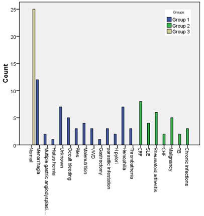
3.2. Underlying etiology of sideropenic anemia
in the study patients
The most prevalent causes of IDA were menorrhagia,
hemophilia, unknown etiology and occult bleeding
while those for ACD were CRF, rheumatoid arthritis,
malignancy and systemic lupus erythematosus in
descending order. Benzidine test was positive
in 9.4 % of patients with IDA denoting occult
blood in stools as in figure 1.
3.3. Splenomegaly in patients with IDA compared
with the sideropenic and healthy control subjects
Splenomegaly was present in 11.3%, 40 % and 0
% of groups 1, 2 & 3, respectively. In IDA
half most of the patients had Hackett's G1 (9.4%)
and only 1.9% had G2, also in ACD G1 comprised
33.3% followed by grade 0 (16.7%) and lastly grade
2 (6.7%). However splenomegaly was sonographically
proven only in 5-patients of group 1 (9.4%), and
10 patients in group 2 (33.3%). Table 1 shows
distribution of splenomegaly in the study groups,
and Table 5 shows diagnostic performance of US
variables in predicting splenomegaly among groups
1 & 2.
Splenomegaly in patients with IDA was commoner
in males 4(80%) and rare in those from urban community
(20%). Their medical history denoted insufficient
dietary intake of iron, and they had pica and
malnutrition. They were excessive drinkers of
tea (100%) with angular stomatitis (80%), and
koilonychias (100%). Hepatomegaly was associated
with splenomegaly in 0 % & 90% of patients
of IDA and sideropenic controls, respectively.
When we correlated hematological parameters with
grading of splenomegaly in patients with IDA they
were negatively associated with HB, MCV, and reticulocytes,
and all patients had severe anemia (Hb ranged
from 3.2-6 g/dl). Table 6 showed factors associated
with splenomegaly in patients with IDA.
The stool analyses showed Giardia lamblia cysts
in 2-patients with IDA and splenomegaly, hookworm
ova in 1 patient and occult bleeding in 1 patient.
The possible etiology of IDA in the other patient
was unknown, however the defective dietary intake
of iron was marked in all patients and was continuous
for many years.
Follow up of patients with SAs and splenomegaly
after treatment revealed gradual progressive reduction
of splenic size with increase in hemoglobin. After
3-months follow up spleen was nearly impalpable
in those with grade 1 Hackett's however dullness
at Traube's area was still detected in patients
with grade 2. Splenomegaly was still sonographically
detected in 2 patients. These findings were noted
in patients with IDA (group 1). On the contrary
splenic size remained stable in the sideropenic
control patients (group 2).
Table 1: Demographic and clinical characteristics
of the study groups (total n = 108)
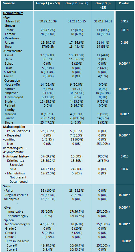
Data were presented as mean± SD, or as
percentage from the total number.
Table 2: Laboratory and hematologic characteristics
of the study groups (total no = 108)
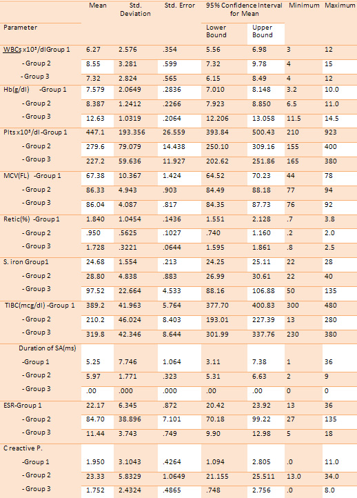
N.B. WBCs= white blood cells, Hb= hemoglobin,
MCV= mean corpuscular volume, Plt= platelet, Retic=
reticulocyte count, ESR= erythrocytic sedimentation
rate, C reactive P= C reactive protein. Duration
of anemia in months. Data were presented as mean±
SD. P value was significant at 0.05 level.
Table 3: Tukey multiple comparison test of
quantitative variables of patients with SAs compared
with the controls (total no=108)
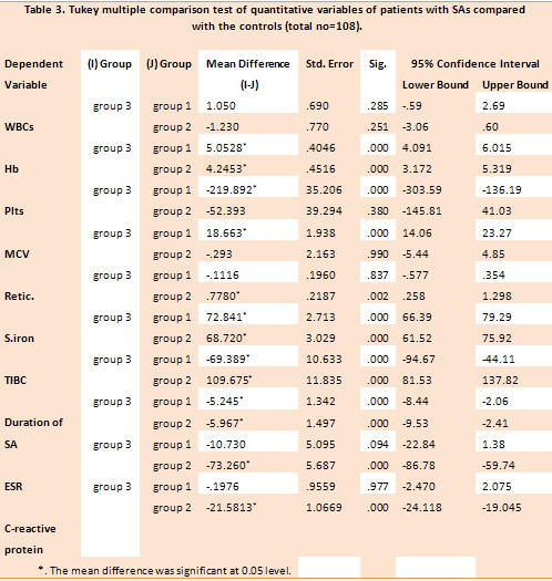
N.B. WBCs= white blood cells x103, Hb= hemoglobin
g/dl , MCV= mean corpuscular volume FL, Plts=
platelets x103, Retic= reticulocyte count%, ESR=
erythrocytic sedimentation rate mm/hr, C reactive
P= C reactive protein mg/dl , TIBC= total iron
binding capacity mcg/dl, S. iron= serum iron.
Table 4: Degree and treatment modalities of
SAs in the study patients (total n = 83)
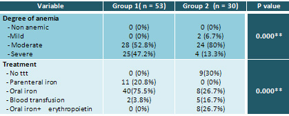
Table 5: Diagnostic performance of US variables
in predicting splenomegaly among groups 1&2
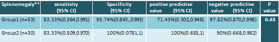
*P value <0.05 , ** No splenomegaly found in
group 3.
Table 6: Factors associated with splenomegaly
in patients with IDA (total no = 53)
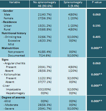
N.B. P value was significant at 0.05 level.
Figure 1: Causes of sideropenic anemias in
the study patients
SAs are hypoproliferative anemias
that are caused by iron deficiency and/or decreased
erythropoietin (EPO) production and/or reduced
response to EPO, the latter due to resistance
of target cells to EPO action or reduced number
of cells. (24) This study was conducted to elucidate
splenomegaly in patients with SAs in terms of
occurrence, clinical and hematological profile
and the effect of treatment on splenomegaly. The
study focused on IDA while ACD was used as a sideropenic
control.
In this study SAs were commoner in females; IDA
was more prevalent in rural residence while urbanization
was obvious in sideropenic controls. Furthermore
IDA was commoner in students and housewives while
ACD in those who did regular office work. These
expected results were explained by increased prevalence
of iron deficiency and chronic inflammatory diseases
in females and higher number of vegans in rural
communities. (25, 26)
This study confirmed a direct relationship between
excessive intake of tea and incidence of IDA.
A common custom in Egypt is to drink nearly 2gms/250
mls of red tea right after each meal particularly
after lunch. Numerous studies reported that tea
hinders iron absorption and advise tea drinkers
to have their cups at least 1 hour after a meal.
(27-29)
Manifestations of tissue iron deficiency were
much higher than that in comparable studies; this
could be explained by the longer duration of iron
deficiency in our patients. However results of
this study were accordant with others in showing
positive association between the degree of tissue
iron deficiency and severity of IDA besides revealing
that angular cheilitis was more prevalent than
koilonychias. Both angular cheilitis and koilonychias
were explained by deficiency of iron based enzymes
in the mucosal and epithelial tissues. (30,31)
In accordance with other studies, reduced Hb,
MCV, MCHC, and thrombocytosis were the CBC features
of IDA; on the contrary normocytosis and normochromia
were evident in sideropenic controls. (32) An
interesting finding was laboratory detection of
mild anemia in asymptomatic controls with the
minimum hemoglobin 11.5g/dl in females. This finding
denoted that the lower cut of value of hemoglobin
should be tailored for each population specifically.
In this study the most common etiology of IDA
was menorrhagia while chronic renal failure was
the commonest cause of ACD. This study confirmed
the findings of others that IDA could retard growth
and development in children and also reaffirmed
that gastrointestinal tract blood loss and H-pylori
infections are common causes of IDA. As reported
by others, hiatus hernia was the underlying etiology
of IDA in one of our patients. (33-36) Although
the recommended daily requirements of iron are
very small, IDA due to ineffective dietary intake
was noted in our patients. This could be explained
by most of them being from a rural community.
Accordingly the main elements of their diets were,
milk and milk products, fruits and vegetables;
both are poor sources of iron.
Splenomegaly was detected in approximately one
tenth of patients with IDA; this was albeit consistent
and inconsistent with other studies. Unlike other
studies there was no detectable splenomegaly in
the control group. (37-39)
When considering the sideropenic control group
splenomegaly was detected in more than half of
the patients and was closely related to the underlying
etiology of anemia, furthermore hepatosplenomegaly
was evident in a considerable proportion of patients.
However there was no association between the degree
of anemia and Hackett's grading of splenomegaly
in the sideropenic control group. As reported
by others, treatment of the underlying etiology
of anemia improved hematological profile of the
patient, (18,40,41) however it did not affect
splenic size. This denoted that the etiopathogenesis
of splenomegaly in the sideropenic controls is
not related to the anemia itself.
In this study patients with IDA and splenomegaly
were mostly from a rural community and the vast
majority of them were males in their late teens
or early twenties. Their nutritional history denoted
ineffective dietary supply of iron and pica. Parasitic
infestation was detected in 3 of the patients.
They were suffering from IDA for years, with periods
of interrupted iron supplementation. Concomitant
with other studies splenomegaly was impalpable
or mild to moderate (Hackett's grades 1 &
2) in most of the patients. Furthermore the degree
of splenomegaly was positively correlated with
the severity of anemia. This was consistent with
Hussain et al and inconsistent with Dabadghao
and his coworkers. (14, 42,43) Notably this study
showed reduction in splenic size with correction
of hemoglobin. Another important finding was the
strong association between splenomegaly and kioilonychia
in patients with IDA; kioilonychia is a sign of
severe long standing IDA.(30) Expectedly the degree
of splenomegaly will be directly correlated with
both the severity and duration of IDA; this assumption
was proved with the results of the current study.
The etiopathogenesis of splenomegaly in patients
with IDA is still unclear. However the most acceptable
explanation is that IDA is a hypoproliferative
anemia with poikilocytosis and anisocytosis both
leading to splenic hyperplasia. Another explanation
is extramedullary hematopoiesis. This could in
turn explains the rare association of hepatomegaly
and splenomegaly in patients with IDA. The latter
assumption could explain the association of splenomegaly
with duration and severity of IDA, furthermore
it explained resolution of splenomegaly with correction
of ID. Although malnutrition was detected in our
patients it could not explain the occurrence of
splenomegaly in IDA as it was a micronutrient
malnutrition rather than a protein energy malnutrition.(
44) However the presence of giardiasis in 2 of
our patients could be a contributing factor for
development of splenomegaly in them. (25)
In conclusion the current study
demonstrated that splenomegaly had considerable
clinicohematologic significance in patients with
SAs. Hackett's grade 1, or 2 splenomegaly was
a common finding in patients with severe, chronic
IDA that was mainly caused by malnutrition. Furthermore
iron supplementation and replenishment of iron
stores led to gradual resolution of splenomegaly.
This denoted that splenomegaly, in patients with
IDA, is a very simple clinically based diagnostic/prognostic
index. However every effort has to be made to
exclude thalassemia in patients with microcytic
hypochromic anemia and splenomegaly before prescribing
iron supplementation. This was not the case in
patients with ACD where splenomegaly was found
to be related to the underlying etiology of anemia
rather than the anemia itself.
Based on the findings of this study we recommended
reducing tea drinking particularly in children
and females who are already in a state of negative
iron balance due to increased demands. Furthermore
we advise people to avoid drinking tea after the
main meal where a great proportion of iron requirements
are supplied.
Acknowledgements
The authors wish to express deep thanks and gratitude
to the entire control group who volunteered participation
in the study for the seeking of robust medical
knowledge. Also great thanks to hematology residents
and paramedical staff at Assiut University Hospital,
Assiut, Egypt for their help during data collection.
[1]- Cavill I., Auerbach
M., Bailie G.R., Barrett-Lee P., Beguin Y., Kaltwasser
P., et al. Iron and the anaemia of chronic disease:
a review and strategic recommendations. Current
Medical Research and Opinion 2006; 22: 731-737.
[2]- Dallman PR, Yi PR, Johnson C. Prevalence
and causes of anemia in the United States, 1976
to1980. Am J Clin Nutr 1984; 39:437-45.
[3]- WHO Global Database on Anemia.. Worldwide
Prevalence of anemia 1993-2005. Geneva, Switzerland:
World Health Organization, 2008.
[4]- Alvarez-Herna´ndez, X., Lice´aga,
J., McKay, I.C. & Brock, J.H. Induction of
hypoferremia and modulation of macrophage iron
metabolism by tumor necrosis factor. Laboratory
Investigation 1989 ; 61: 319-322.
[5]- Cazzola M., Ponchio L., de Benedetti F.,
Ravelli A., Rosti V., Beguin, Y., et al. Defective
iron supply for erythropoiesis and adequate endogenous
erythropoietin production in the anemia associated
with systemic-onset juvenile chronic arthritis.
Blood 1996; 87: 4824-4830.
[6]- Ferrucci L., Semba R.D., Guralnik J.M., Ershler
W.B., Bandinelli, S., Patel K.V., et al. Proinflammatory
state, hepcidin, and anemia in older persons.
Blood 2010; 115: 3810-3816.
[7]- Ganz T, Nemeth E. Hepcidin and disorders
of iron metabolism. Annu Rev Med. 2011; 62:347-360.
[8]- Busbridge M., Griffiths C., Ashby D., Gale
D., Jayantha, A., Sanwaiya A. et al. Development
of a novel immunoassay for the iron regulatory
peptide hepcidin. British Journal of Biomedical
Science 2009; 66: 150-157.
[9]- Skikne BS, Punnonen K, Caldron PH, Bennett
MT , Rehu M, Gasior GH, et al. Improved differential
diagnosis of anemia of chronic disease and iron
deficiency anemia: a prospective multicenter evaluation
of soluble transferring receptor and the sTfR/log
ferritin index. Am J Hematol. 2011; 86(11):923-927.
[10]- Brasse-Lagnel C., Karim Z., Letteron P.,
Bekri S., ado A. & Beaumont C. Intestinal
DMT1 cotransporter is down-regulated by hepcidin
via proteasome-internalization and degradation.
Gastroenterology 2011; 140: 1261-1271.
[11]-Hoffman RM, Jaffe PE. Plummer-Vinson syndrome.
A case report and literature review. Arch Intern
Med. 1995; 155(8):2008-11.
[12]- Prasad AS, Halsted JA, Nadimi M. Syndrome
of iron deficiency anemia, hepatosplenomegaly,
hypogonadism, dwarfism and geophagia. Am J Med
1961; 31:532-46.
[13]-King R, Mizban N, Rajeswaran C. Iron deficiency
anaemia due to hypopituitarism. Endocrine Abstr
2009; 19:278.
[14]- Safaa A. A. Khaled. Aggravation of Iron
Deficiency Anemia after Hormone Replacement Therapy
in a Patient with Hypopituitarism and Hepatosplenomegaly
. Int. J. Adv. Res. Biol. Sci. 2015; 2(12):324-329.
[15]- Shah A. Iron deficiency anemia Part II (Etiopathogenesis
and diagnosis). Indian J Med Sci 2004? 58:134-7.
[16]- Ogilvie C. Evans CC: Splenomegaly. In: Wratt
G. Symptoms and signs in Tropical Disease. Chamberlaine's
symptoms and signs in Clinical medicine: 12th
edition. Butterworth- Heineman, 1997, ( 315-324).
[17]- Benter T, Klühs L, Teichgräber
U. Sonography of the spleen. J Ultrasound Med.
2011; 30(9):1281-93.
[18]- Doyle M.K., Rahman M.U., Han C., Han J.,
Giles J., Bingham C.O. et al. Treatment with infliximab
plus methotrexate improves anemia in patients
with rheumatoid arthritis independent of improvement
in other clinical outcome measures-a pooled analysis
from three large, multicenter, double-blind, randomized
clinical trials. Seminars in Arthritis and Rheumatism
2009; 39:123-131.
[19]- Liu K, Kaffes AJ. Iron deficiency anemia
: a review of diagnosis, investigation and management.
Eur J Gastroenterol Hepatol. 2012; 24(2):109-116.
[20]- Goddard AF, James MW, Mclntyre AS, Scott
BB. British Society of Gastroenterology Guidelines
for the management of iron deficiency anemia.
Gut. 2011; 60 (10): 1309-1316.
[21]- Auerbach M., Ballard H., Trout J.R., McIlwain
M., Ackerman A., Bahrain H., et al. Intravenous
iron optimizes the response to recombinant human
erythropoietin in cancer patients with chemotherapy-related
anemia: a multi-center, open-label, randomized
trial. Journal of Clinical Oncology 2004; 22:1301-1307.
[22]- Auerbach, M. & Ballard, H. Clinical
use of intravenous iron: administration, efficacy,
and safety. Hematology. American Society of Hematology.
Education Program, 2010; 338-347.
[23]- World Medical Association. World Medical
Association Declaration of Helsinki Ethical Principles
for Medical Research Involving Human Subjects.
Clinical Review & Education 2013; 310(20):
2191-94.
[24]- Faquin W.C., Schneider T.J. & Goldberg
M.A. Effect of inflammatory cytokines on hypoxia-
induced erythropoietin production. Blood 1992;
79: 1987-1994.
[25]- Kim JY, Shin S, Han K, Lee KC, Kim JH, Choi
YS, et al. Relationship between socioeconomic
status and anemia prevalence in adolescent girls
based on the fourth and fifth Korea National Health
and Nutrition Examination Surveys. Eur J Clin
Nutr. 2014 ; 68(2):253-8.
[26]- Betsy Lozoff, M.D, Elias Jimenez, and Julia
B. Smith, Double burden of iron deficiency in
infancy and low socio-economic status: a longitudinal
analysis of cognitive test scores to 19 years.
Arch Pediatr Adolesc Med. 2006 ; 160(11): 1108-1113.
[27]-Nelson M. , Poulter J. Impact of tea drinking
on iron status in the UK: a review. J Hum Nutr
Dietet 2004; 17: 43-54.
[28]-Disler PB, Lynch SR, Charlton RW, Torrance
J D, Bothwell T H, Walker R B, et al. The effect
of tea on iron absorption. Gut 1975; 16:193-200.
[29]- J P Kaltwassera, ,E Wernerb,, K Schalka,
C Hansenb, R Gottschalka, C Seidl. Clinical trial
on the effect of regular tea drinking on iron
accumulation in genetic haemochromatosis. Gut
1998; 43:699-704 doi:10.1136/gut.43.5.699.
[30]- Kumar G, Vaidyanathan L, Stead LG. Images
in emergency medicine. Koilonychia, or spoon-shaped
nails, is generally associated with iron-deficiency
anemia. Ann Emerg Med. 2007; 49(2):243, 250.
[31]- Uchida T, Matsuno M, Ide M, Kawachi Y. The
frequency and development of tissue iron deficiency
in 6 iron deficiency anemia patients with Plummer-Vinson
syndrome. Rinsho Ketsueki. 1998 39(11):1099-102.
Japanese
[32]- Yates JM, Logan EC, Stewart RM. Iron deficiency
anemia in general practice: clinical outcomes
over three years and factors influencing diagnostic
investigations. Postgraduate Medical Journal.
2004; 80(945): 405-410.
[33]- Bandhu R, Shankar N, Tandon OP. Effect of
iron on growth in iron deficient anemic school
going children. Indian J Physiol Pharmacol. 2003;
47:59-66.
[34]- Soliman AT, Al Dabbagh MM, Habboub AH, Adel
A, Humaidy NA, Abushahin A. Linear growth in children
with iron deficiency anemia before and after treatment.
J Trop Pediatr. 2009; 55:324-7.
[35]- Rockey DC, Cello JP. Evaluation of the gastrointestinal
tract in patients with iron deficiency anemia.
New England Journal of Medicine 1993; 329: 1691-5.
[36]- Windsor CW, Collis JL. Anaemia and hiatus
hernia: experience in 450 patients. Thorax 1967;
22(1): 73-8.
[37]- Nadeem A., Ali N., Hussain T. Anwar M. Frequency
and etiology of splenomegaly in adults seeking
medical in Combined Military Hospital, Attock.
J Ayub Med Coll 2004; 16(4): 44-47.
[38]-Sundaresan JB, Dutta TK, Badrinath S , Jagdish
S, Basu D. A Hospital based Study of Splenomegaly
with special reference to the Group of Indeterminate
Origin. J Indian Med Assoc 2008; 106(3):150- 152.
[39]-McIntyre OR, Ebaugh FG. Palpable spleen in
college freshmen. Ann Intern Med 1967? 66: 30-33.
[40]-Ali N, Anwar M, Ayyub A, Nadeem M, Ejaz A,
Qureshi AH, et al. Hematological evaluation of
splenomegaly. J Coll Physicians Surg Pakistan
2004; 14(7):404-406.
[41]- Papadaki, H.A., Kritikos, H.D., Balatas,
V., Boumpas, D.T. & Eliopoulos, G.D. Anemia
of chronic disease in rheumatoid arthritis is
associated with increased apoptosis of bone marrow
erythroid cells: improvement following antitumor
necrosis factor-alpha antibody therapy. Blood,
2002; 100: 474-482.
[42]-Hussain I, Ahmed I, Mohsin A. Causes of splenomegaly
in adult local population presenting at tertiary
care centre in Lahore. Pakistan J Gastroenterol
2002; 16(1):12-16.
[43]- Dabadghao VS, Diwan AG, Raska AM. A Clinicohaematological
Profile of Splenomegaly. Bombay Hospital Journal
2012; 54:10-17.
[44]- Anwar, I. and J.A. Awan. Nutritional status
comparison of rural with urban school children
in
Faisalabad, Pakistan. J. Rural Remote Health,
, 2003 1-7.
[45]-Charalabopoulos K, Charalabopoulos A, Papadopoulou
CH, Papalimneou V. Giardia lamblia intestinalis:
a new pathogen with possible link to Kikuchi-Fujimoto
disease. An additional element in the disease
jigsaw. Int J Clin Pract. 2004 Dec;58(12):1180-3.
|